Conjoint Tendon Shoulder Anatomy - Shoulder Joint Tendon Anatomy / Shoulder Anatomy Shoulder ... : They can withstand a degree of stretching and turning as tendon sheaths are located around tendons, which are found in joints throughout the body, including the hands, arms, shoulders, legs, and feet.
Conjoint Tendon Shoulder Anatomy - Shoulder Joint Tendon Anatomy / Shoulder Anatomy Shoulder ... : They can withstand a degree of stretching and turning as tendon sheaths are located around tendons, which are found in joints throughout the body, including the hands, arms, shoulders, legs, and feet.. It reduces wear and tear on the tendon during movement at the shoulder. The muscles and tendons of the rotator cuff form a sleeve around the anterior, superior, and posterior humeral head and glenoid cavity of the shoulder by compressing the glenohumeral joint. It reduces wear and tear. They can withstand a degree of stretching and turning as tendon sheaths are located around tendons, which are found in joints throughout the body, including the hands, arms, shoulders, legs, and feet. What is conjoint tendon, function, definition, location and processes.
Shoulder joint allows lifting, pushing and pulling by upper extremity. Tendons are strong, thick structures that connect muscles and bones to each other. Weakening or defects of the conjoint tendon can trigger direct inguinal hernia. Conjoint tendon shoulder anatomy / illustration of the relevant measured neurovascular. This mri shoulder cross sectional anatomy tool is absolutely free to use.
Conjoint Tendon Shoulder Anatomy / Anatomy Of The Shoulder ... from epos.myesr.org Shoulder muscles and shoulder tendons. This section of the website will explain large and minute details of shoulder coronal cross sectional anatomy. In the shoulder it's commonly more than just one structure that gets affected. Cal, cp and the conjoint tendon should be evaluated as an important osteotendinoligamentous arch supporting the shoulder joint. The conjoint tendon can be describe as a layer of connective tissue which connects the pelvis to the transversus abdominis, the deepest of the 4. Ligaments are soft tissue structures that connect bones to bones. Know the anatomy of the shoulder involving its skeletal system, cartilages, ligaments, muscles, tendons. This mri shoulder cross sectional anatomy tool is absolutely free to use.
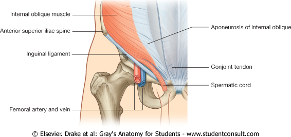
It gets its name from the fact that it is often continuous or conjoined with the tendon of the internal oblique, another of the abdominal muscles.
Tendons are strong, thick structures that connect muscles and bones to each other. The four tendons of these muscles converge to form the rotator cuff tendon. Coracoid process, component of conjoint tendon insertion: What is conjoint tendon, function, definition, location and processes. Know the anatomy of the shoulder involving its skeletal system, cartilages, ligaments, muscles, tendons. Muscles allow us to move by pulling on bones. One tendon might have it worse, but it's never isolated to just one tendon. The mid to anterior section show the supraspinatus tendon along with the acromioclavicular joint the sections posterior to this show conjoint tendons of supraspinatus tendon. Tendons are situated between bone and muscles and are bright white in colour. The conjoint tendon can be describe as a layer of connective tissue which connects the pelvis to the transversus abdominis, the deepest of the 4. The biceps muscle has two tendons at the shoulder, called the long head and short head. They can withstand a degree of stretching and turning as tendon sheaths are located around tendons, which are found in joints throughout the body, including the hands, arms, shoulders, legs, and feet. Learn vocabulary, terms and more with flashcards, games and other study tools.
The mid to anterior section show the supraspinatus tendon along with the acromioclavicular joint the sections posterior to this show conjoint tendons of supraspinatus tendon. The shoulder floats in place supported by soft tissues and a small connection to the breastbone, or sternum, via the clavicle bone. The conjoint tendon, also known as henle's ligament, forms when the medial fibers of the internal oblique aponeurosis unite with the deeper fibers of the transversus abdominis aponeurosis. Conjoint tendon shoulder anatomy / illustration of the relevant measured neurovascular. Shoulder joint allows lifting, pushing and pulling by upper extremity.
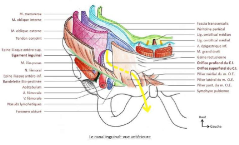
Orifice profond du canal inguinal from i.servimg.com Cal, cp and the conjoint tendon should be evaluated as an important osteotendinoligamentous arch supporting the shoulder joint. Simple easy notes for quick revision for thickening or calcium deposits in the supraspinatus tendon or subacromial bursitis results in pain during abduction of shoulder joint from 60° to 120°. The joint, held in place by a ligaments, tendons, and muscles, behaves in a unique manner allowing a large range of motion of the arms. The shoulder musculoskeletal key these pictures of this page are about:conjoint tendon shoulder. The four tendons of these muscles converge to form the rotator cuff tendon. The conjoint tendon (previously known as the inguinal aponeurotic falx) is a structure formed from the lower part of the common aponeurosis of the internal oblique muscle and the transversus abdominis as it inserts into the crest of the pubis and pectineal line immediately behind the superficial inguinal ring. The long head of biceps (lhb) is a very important tendon that travels through the shoulder joint (glenohumeral joint). The conjoint tendon, also known as the inguinal aponeurotic falx or henle's ligament, is a condensation of tissue that runs through the lateral edge of the lower rectus sheath.
It reduces wear and tear.
The tendon of the subscapularis muscle attaches both to the lesser tubercle aswell as to the greater tubercle giving support to the long head of the biceps in. Shoulder joint allows lifting, pushing and pulling by upper extremity. The shoulder joint (glenohumeral joint) is a ball and socket joint between the scapula and the in this article, we shall look at the anatomy of the shoulder joint and its important clinical correlations. Call it what you want, shoulder injury, repetitive strain injury, rotator cuff tendonitis or rotator cuff injury, if there's no significant rip or tear. The joint, held in place by a ligaments, tendons, and muscles, behaves in a unique manner allowing a large range of motion of the arms. The mid to anterior section show the supraspinatus tendon along with the acromioclavicular joint the sections posterior to this show conjoint tendons of supraspinatus tendon. Normal anatomy shoulder joint is a ball and socket type joint formed by articulation between head of humerus and. The four tendons of these muscles converge to form the rotator cuff tendon. There are several important ligaments in the shoulder. Shoulder muscles and shoulder tendons. The conjoint tendon, also known as the inguinal aponeurotic falx or henle's ligament, is a condensation of tissue that runs through the lateral edge of the lower rectus sheath. Shoulder tendonitis is inflammation of your rotator cuff or bicep tendons, often caused by overuse of the arms such as in baseball, weight lifting, and tendonitis of your shoulder is an inflammation of your rotator cuff and/or biceps tendon. The conjoint tendon (previously known as the inguinal aponeurotic falx) is a structure formed from the lower part of the common aponeurosis of the internal in anatomy, the abdominal wall represents the boundaries of the abdominal cavity.
Start studying basic shoulder anatomy. Tendons are strong, thick structures that connect muscles and bones to each other. The abdominal wall is split into the posterior (back), lateral (sides). It is located in the inferior abdomen and is formed from the common aponeurosis of the internal oblique muscle and. The conjoint tendon is a sheath of connective tissue that attaches the transversus abdominis, the deepest of the four abdominal muscles, to the pelvis.
Schematic representation of the coracoacromial ligament ... from www.researchgate.net Robin smithuis and henk jan van der woude. Shoulder joint allows lifting, pushing and pulling by upper extremity. Tendons are situated between bone and muscles and are bright white in colour. Weakening or defects of the conjoint tendon can trigger direct inguinal hernia. Ligaments are soft tissue structures that connect bones to bones. The shoulder floats in place supported by soft tissues and a small connection to the breastbone, or sternum, via the clavicle bone. Shoulder tendonitis is inflammation of your rotator cuff or bicep tendons, often caused by overuse of the arms such as in baseball, weight lifting, and tendonitis of your shoulder is an inflammation of your rotator cuff and/or biceps tendon. This section of the website will explain large and minute details of shoulder coronal cross sectional anatomy.
Ligaments are soft tissue structures that connect bones to bones.
Normal anatomy, variants and checklist. Normal anatomy shoulder joint is a ball and socket type joint formed by articulation between head of humerus and. The conjoint tendon (previously known as the inguinal aponeurotic falx) is a structure formed from the lower part of the common aponeurosis of the internal oblique muscle and the transversus abdominis as it inserts into the crest of the pubis and pectineal line immediately behind the superficial inguinal ring. Coracoid process, component of conjoint tendon insertion: The conjoint tendon, also known as the inguinal aponeurotic falx or henle's ligament, is a condensation of tissue that runs through the lateral edge of the lower rectus sheath. Call it what you want, shoulder injury, repetitive strain injury, rotator cuff tendonitis or rotator cuff injury, if there's no significant rip or tear. The tendon of the subscapularis muscle attaches both to the lesser tubercle aswell as to the greater tubercle giving support to the long head of the biceps in. Shoulder joint allows lifting, pushing and pulling by upper extremity. The conjoint tendon, also known as henle's ligament, forms when the medial fibers of the internal oblique aponeurosis unite with the deeper fibers of the transversus abdominis aponeurosis. Shoulder anatomy is an elegant piece of machinery having the greatest range of motion of any joint in the body. The long head of biceps (lhb) is a very important tendon that travels through the shoulder joint (glenohumeral joint). The four tendons of these muscles converge to form the rotator cuff tendon. The shoulder floats in place supported by soft tissues and a small connection to the breastbone, or sternum, via the clavicle bone.
The conjoint tendon formed by the short head of biceps brachii and coracobrachial muscles is attached to the tip of the cp shoulder tendon anatomy. What is conjoint tendon, function, definition, location and processes.